
 Faculty of Science, Technology and Medicine
 Faculty of Law, Economics and Finance
 Faculty of Humanities, Education and Social Sciences
 Interdisciplinary Centre for Security, Reliability and Trust
 Luxembourg Centre for Systems Biomedicine
 Luxembourg Centre for Contemporary and Digital History
 Luxembourg Centre for European Law
 Luxembourg Centre for Socio-Environmental Systems
Our research
To study the underlying mechanisms of Parkinson’s disease (PD), our team employs molecular, ‘omics’ and single-cell approaches. We work with both post-mortem human midbrain samples and cell cultures derived from induced pluripotent stem cells (iPSC). These two models allow us to explore different aspects of the disease.
Some of our research projects focus on the role of mitochondria in neurodegeneration. Others investigate the composition of the midbrain and the interactions between neurons and other cell types, such as microglia and astrocytes. Specifically, we are interested in energy metabolism and inflammatory signalling as processes that accelerate neuronal demise in Parkinson’s disease patients.
The Molecular and Functional Neurobiology group was established under the auspices of the Luxembourg National Research Fund’s ATTRACT Programme.
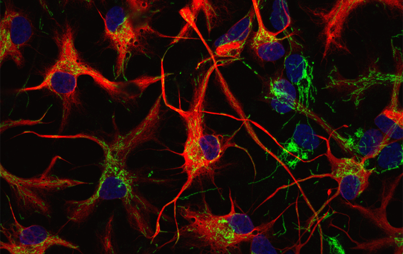
Figure: Labelling of the mitochondrial network (green) in iPSC-derived PD patient astrocytes (red).
Our research projects
The Molecular & Functional Neurobiology group is involved in several research projects focusing on Parkinson’s disease:
-
Duration:
-
Funding source:
Luxembourg National Research Fund (FNR) ATTRACT project Model-IPD
-
Researchers:
-
Partners:
collaboration with two other research groups from the LCSB
-
Description:
Idiopathic Parkinson’s disease (IPD) is a complex condition, in which a genetic cause has not been defined, and thus encompasses patients with diverse underlying disease mechanisms. Mitochondrial dysfunction was implicated in PD leading to impaired energy metabolism in dopaminergic neurons of the substantia nigra. In this project, we are assessing the value of mitochondrial-based stratification approaches to help disentangle the complexity of PD. Our study aims at identifying approaches to categorise IPD patients into homogeneous subgroups that will facilitate mechanistic studies, and drug screening ventures.
In collaboration with the Biomedical Data Science group, we have explored published genetic and functional datasets from IPD samples of the FOUNDIN-PD study. RNA-seq data of iPSC-derived 65d-old dopaminergic neurons show that the abundance of a gene associated to familial PD can be used to cluster IPD patients into two subgroups, which present distinctive mitochondrial pathways signatures. Identified altered pathways are being explored in an independent cohort comprising iPSCs from seven IPD cases and four age-matched controls. We are investigating parameters such as mtDNA maintenance, mitochondrial morphology and mitophagy in 2D neuronal cultures. Cluster and machine learning analyses of these results identified a subclass of three IPD patients depicting aberrant mitochondrial function. Together with the Developmental & Cellular Biology group, we are exploring the implication of our findings in more complex models such as midbrain organoids, generated from the same cohort. We are evaluating cell type dynamics and viability, neuronal development and function as well as astrocyte function. Based on our findings, we are investigating possible links between mitochondrial dysfunction and astroglial activation.
-
Project details (PDF):
-
Duration:
-
Funding source:
Luxembourg National Research Fund (FNR) ATTRACT project Model-IPD
-
Researchers:
-
Partners:
-
Description:
To date, transcriptomic studies in post-mortem human midbrain were based on bulk RNA sequencing technologies, hindering the study of the contribution of individual cell types to disease pathology. To overcome this challenge, we performed single-cell RNA sequencing of post-mortem human midbrain tissue of five IPD patients and six age-matched healthy controls to obtain an unbiased and global view of the cell type composition and cellular phenotypes of IPD. We identified all major cell types in the midbrain including different neuronal subtypes, glia and microvasculature cells and detected differentially expressed genes in each of those cell types. Moreover, we discovered an IPD-specific neuronal cluster characterized by the overexpression of CADPS2 and low TH expression, which suggests dysfunctional dopaminergic neurons. In addition, we observed a disease-specific upregulation of microglia and astrocytes further strengthening the role of neuroinflammation in PD (Smajic et al., Brain, 2021). With additional analyses, we demonstrated the contribution of neuromelanin to the microglia activation in PD.
-
Project details (PDF):
-
Duration:
-
Funding source:
Luxembourg National Research Fund (FNR) CORE Junior project NeuroFlame
-
Researchers:
-
Partners:
-
Description:
“Neuroinflammation has been a hallmark of PD since the early 1980s. Many studies in human serum and cerebrospinal fluid samples have shown a significant upregulation of proinflammatory cytokines and chemokines in PD patients. This includes our own work, which identified increased levels of circulating cell-free mtDNA and the proinflammatory cytokine IL-6 in blood serum from PD patients with PRKN or PINK1 mutations (Borsche et al., Brain, 2020). Inspired by these biomarker studies, we sought to investigate, if IPD inflammation can be modeled in iPSC-derived microglia. First, by analysing multiple publicly available bulk RNAseq and snRNAseq datasets from post-mortem midbrain tissue, we discovered a significant upregulation of IL1B and IL10 in IPD patients compared to healthy controls. Furthermore, we identified microglia as being the predominant cell type involved in the propagation of inflammatory cascades. Second, to validate these findings in vitro, we generated iPSC-derived microglia. By treating our control and IPD iPSC-derived microglia with LPS, we indeed observed a higher expression of both IL1B and IL10 in IPD microglia. Moreover, this upregulation coincided with elevated levels of the NLRP3 inflammasome, indicative of stronger immune priming in IPD microglia (Badanjak et al., Front Cell Dev Biol, 2021).
Alpha-synuclein is a key protein involved in idiopathic as well as genetic forms of PD, which has been associated with the release of inflammatory mediators and mitochondrial dysfunction. Using iPSC-derived microglia as well as neuron-glia co-cultures, we aim at studying the effect of alpha-synuclein on mitochondria modulation and NLRP3 inflammasome activation.”
-
Project details (PDF):
-
Duration:
-
Funding source:
co-funded by the Luxembourg National Research Fund (FNR) and the DFG
-
Researchers:
-
Partners:
This project is part of the Research Unit “ProtectMove.
-
Description:
The manifestation of PD and the age of onset are not exclusively determined by the mutation identity, for instance in the LRRK2 gene. So far, only few factors have emerged, which define disease penetrance or constitute signs of advanced progression in LRRK2-PD. By contrast, in fibroblasts from manifesting and non-manifesting G2019S mutation carriers, we detected a correlation between mtDNA deletions and disease status (Ouzren et al., Ann Neurol, 2019). Furthermore, cells from manifesting G2019S carriers had impaired complex I function as well as increased mitochondrial mass and mtDNA copy number, suggestive of impaired mitophagy. Finally, elevated expression of Nrf2 implicates ROS scavenging in the penetrance of LRRK2-PD (Delcambre et al., Front Neurol, 2020). Thus, we now speculate that, in manifesting G2019S mutation carriers, increased LRRK2 kinase activity interferes with Nrf2 antioxidant signaling, which in turn (i) promotes mtDNA damage, (ii) mediates ccf-mtDNA release, and eventually (iii) triggers pro-inflammatory signaling. We are exploring this hypothesis in patient iPSC-derived neurons and microglia. Of note, iPSC-derived microglia from G2019S mutation carriers exhibit higher levels of pS1292 LRRK2, a known pathogenic form of phosphorylated LRRK2, and these levels are accompanied by significantly higher levels of pRab10, a known LRRK2 target. Our findings further strengthen the role of LRRK2 kinase activity in the pathogenesis of PD. We are currently investigating the therapeutic kinase inhibitor MLi-2, with or without inflammatory stimuli, to study the role of kinase activity in the inflammatory processes. In addition, we investigated how environmental toxins may interfere with this molecular mechanism (Lüth et al., Front Aging Neurosci, 2020).
-
Project details (PDF):
-
Duration:
-
Funding source:
Luxembourg National Research Fund (FNR) CAMeSyn,
-
Researchers:
-
Partners:
Dr Johannes Meiser (LIH) and Prof Rejko Krüger (LCSB, LIH). We are also applying a differentiation protocol developed in the group of Prof Jari Koistinaho (University of Helsinki).
-
Description:
Recently, the neurocentric view of PD has been challenged with evidences that the disrupted interaction between neurons and glia contribute to the disease progression. Notably, there is a strong body of evidence indicating that astrocytic metabolism is pivotal to ensure proper neuronal function. In light of these findings, we aim to study the neuronal-astrocytic cooperation in astrocytes derived from patients harboring mutations in SNCA, with a special focus on metabolic alterations that could exacerbated neuronal pathology. Furthermore, we are generating pure non-activated astrocyte cultures from patients and controls, which are suitable for metabolomics studies, as well as cytokine profiling in the context of neuroinflammation.
-
Project details (PDF):
-
Duration:
-
Funding source:
-
Researchers:
-
Partners:
-
Description:
Mutations in Parkin explain up to three quarters of early-onset familial PD cases. Regarding the protein’s function, there is strong evidence that Parkin is involved in the clearance of depolarized mitochondria. Interestingly, recent studies in PRKN knockout mtDNA “mutator” mice also suggested a role for Parkin in the regulation of mitochondrial DNA (mtDNA) homeostasis and cGas/STING signaling which is triggered by mtDNA released into the cytosol. We used PRKN-PD patient-derived blood samples, neurons and microglia to explore this link at the endogenous level. Investigating mitochondrial maintenance pathways, mtDNA integrity, mitochondrial metabolism and cGas/STING-mediated inflammation in these models, we showed that mtDNA disintegration and release is due to a metabolic shift that is sensed by SIRT1, which simultaneously controls mitochondrial biogenesis and clearance. In addition, single-cell mtDNA and RNAseq analyses of postmortem midbrain tissue from a PD patient with PRKN mutations revealed mtDNA somatic mutation accumulation in dopaminergic neurons and an upregulation of microglia. Finally, PRKN-mutant iPSC-derived neuron-microglia co-cultures elicited an enhanced immune response when exposed to mtDNA/LPS. Taken together, our findings suggest that Parkin co-regulates mitophagy, mitochondrial biogenesis and mtDNA maintenance pathways, thereby protecting midbrain neurons from neurodegeneration and -inflammation (Borsche et al. Brain 2020; Wasner et al. Mov Disord 2022; Trinh et al. Brain 2023).
-
Project details (PDF):